
Digital Radiography for Accurate Diagnosis
Digital X-rays are an innovative diagnostic tool that provides a clearer, more detailed view of your teeth and oral structures. Unlike traditional radiographic film, digital radiography uses an electronic sensor to capture images, which are then instantly uploaded to a computer. This technology allows the team at Pearl Dental Care in Florence, KY to closely examine your teeth and surrounding tissues for issues that may not be visible during a routine exam.
With digital X-rays, images can be enlarged and enhanced for better diagnostic accuracy. The advanced software also enables precise measurement of cavities, restorations, and other dental concerns, helping us deliver more accurate and personalized treatment plans.
What Can Digital X-rays Reveal?
At Pearl Dental Care in Florence, KY, digital X-rays are a valuable diagnostic tool that help us detect a wide range of dental issues that may not be visible during a standard examination. These high-resolution images can reveal:
- Bone loss
- Tooth decay, especially in areas between the teeth
- Malignant and benign tumors
- Developmental abnormalities in children
- Cysts and abscesses
- Problems hidden below the gum line
- Internal tooth abnormalities
- Poor root alignment and tooth positioning
With this advanced imaging technology, we’re able to catch potential problems early and create more effective, personalized treatment plans for your long-term oral health.
Are Digital X-Rays Safe?
At Pearl Dental Care in Florence, KY, your safety is always a top priority. While traditional dental X-rays already emit a low level of radiation, digital X-rays reduce that exposure by 80 to 90%, making them an even safer option for patients of all ages.
In fact, the radiation from dental X-rays is comparable to the background radiation we encounter daily—from sunlight, living in a brick home, sharing a bed, or even reading a book for a few hours. The minimal exposure involved in dental imaging should not be a cause for concern.
One of the greatest advantages of digital X-rays is the enhanced diagnostic capability. Images can be enlarged and analyzed with precision, allowing our team to detect abnormalities and measure problem areas with greater accuracy. This advanced technology helps us identify issues early—often before symptoms appear—so we can prevent the need for more invasive or costly treatments later on.
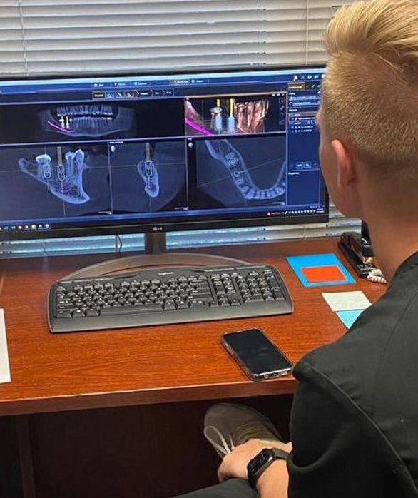
Advanced Imaging with CBCT Technology
In addition to traditional digital X-rays, our office is equipped with CBCT (Cone Beam Computed Tomography)—a powerful 3D imaging tool that provides detailed views of the teeth, jawbone, nerves, and surrounding structures.
CBCT plays a critical role in several areas of care, including:
Dental Implant Planning – Ensures precise placement by mapping bone density, nerve locations, and sinus proximity.
Infection Detection – Helps identify abscesses, bone loss, and other signs of infection that may not be visible on standard X-rays.
Endodontic (Root Canal) Therapy – Offers a clear view of root canals and surrounding anatomy, improving accuracy and treatment outcomes.
This advanced technology allows us to diagnose more effectively, plan with confidence, and provide care that’s safer, more efficient, and tailored to your unique needs.